Medical Ablation Technology developed by MedWaves

Founded in 1998, MedWaves has extensive experience using microwave technologies to solve biomedical problems. MedWaves is the first company to use temperature- and frequency-based feedback mechanisms to enable precise and safe control over the delivery of microwave energy in biomedical applications. MedWaves’ proprietary microwave platform technology provides means for efficient therapeutic energy delivery that are safe, effective, economical and user-friendly. The company’s technology requires no irrigation, no cooling and no return pad like the other products in the market. The following list describes some of the unique microwave ablation technology developed by MedWaves:

  • Dynamic Frequency Selection
  • Efficient Energy Delivery
  • Real-Time Data Feedback
  • Predictable Ablation Zones

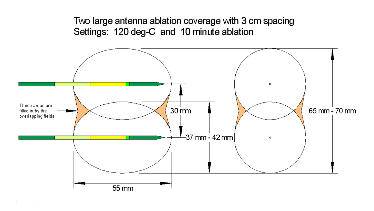
In addition, the MedWaves proprietary microwave platform technology enables you to customize the Ablation Zones to enable precise and safe control over the delivery of energy. The diagrams below describe four (4) different configurations of ablation coverage in terms of spacing, temperature and duration. (Click on each image to zoom in for more detail).

3-large_3-cm_10-min 2-large_3-cm_15-min
2-large_3-cm_10-min 3-large_3-cm_15-min

Examples of MedWaves at Work

The MedWaves AveCure generator and probes have been used in a variety of applications. Please click the links below to see results delivered by MedWaves.

Primary Lung Tumor Microwave Ablation

83-year old female with primary lung tumor, temperature-mode ablation at 110°C and 11 minutes. Below, from left to right:

  • Pre ablation image showing tumor and satellites nearby.
  • During ablation image shows ground-glass effect of microwave coagulation in and around the tumor.
  • Post ablation images at one month follow-up showing scar tissue with no tumor enhancements.

The ablation was performed April 2009 and the patient has no signs of recurrence.

Lung Carcinoid Tumor Microwave Ablation

79-year old female with lung carcinoid, power-mode ablation at 28 watts, 100°C – 100°C and 12 minutes. Below, from left to right:

  • Pre-ablation image showing tumor next to aorta growing front to back and top to bottom.
  • Pre-ablation image shows one of placements of antenna.
  • Post-ablation image showing ground-glass effect of microwave coagulation in and around the tumor.

Post ablation images at six months follow-up showing scar tissue with no tumor enhancements and shrinkage of tissue.

Liver Tumor Microwave Ablation

70-year old male with two colorectal metastasis in the liver, temperature-mode ablation at 110°C. Below, from left to right:

  • Pre-ablation image showing tumor next to the hepatic veins.
  • Post-ablation image showing ablation zone between the veins.
  • Post-one-month-ablation image showing with shrinkage and no enhancements.
  • Pre-ablation image shows smaller tumor more peripherally. Post ablation images showing ellipsoidal zone from 5 minute energy applications

Post ablation one month follow-up showing scar tissue with no enhancement and tissue shrinkage.

Bone Tumor, Osteoid Osteoma Microwave Ablation

13 year old male, right hip Osteoid Osteoma, in pain for several months with a sclerotic rim ablated using minifield antenna, power-mode, 16 watts, 80°C and one minute. Below, from left to right:

  • Pre-ablation MRI T1W and T2W images showing the tumor in right femoral head.
  • Pre-ablation image shows mini field antenna in position for treatment.
  • Pre- and post-ablation dynamic MRI images show complete revascularization of tumor and ablated zone.

Complete pain relief the day after ablation.